- completed
[Cell Mechanosensing]
Identification of gravity-transducers in skeletal muscle cells
- Biology and Biotechnology
ISS Science for Everyone
SCIENCE OBJECTIVES FOR EVERYONE
Identification of gravity-transducers in skeletal muscle cells: Physiological relevance of tension fluctuations in plasma membrane (Cell Mechanosensing) is an investigation that identifies gravity sensors in skeletal muscle cells in order to develop countermeasures to muscle atrophy, a key space health issue. Scientists believe that the lack of mechanical stress from gravity causes tension fluctuations in the plasma membrane of skeletal muscle cells, which changes the expression of key proteins and genes, and allows muscles to atrophy. Muscle cells from rats, and kidney cells from African clawed frogs are tagged with fluorescent gene markers, and attached to an extracellular matrix to study their performance under different tensions, simulating use on earth.
Experiment Description
RESEARCH OVERVIEW
Skeletal muscle is sensitive to mechanical stress. For example, it easily atrophies under microgravity conditions, while exercise stimulates its growth. Several lines of investigation suggest that skeletal muscle cells possess unique sensors for mechanical stress. However, this sensing mechanism of mechanical stress remains unknown. Based on previous studies, it is hypothesized that tension fluctuation in salcolemma (the plasma membrane of skeletal muscle cells) plays an important role in sensing mechanical stress, such as microgravity. Mechanical stress causes changes in tension fluctuation in the salcolemma, resulting in the activation of channels and/or proteases on membrane surface. They transduce intracellular signaling in skeletal muscle cells to express muscle atrophy-associated ubiquitin ligases (atrogenes), including Cbl-b, MAFbx/atrogin-1, and MuRF-1. Investigators have already reported two candidate transducers, MSCL and MSPL. The former is a mechanosensitive channel with a large pore (MSCL) which induces a Calcium Ion (Ca2+) influx. The latter is Mosaic Serine Protease, large form (MSPL) which mediates the shedding of bioactive membrane molecules. In this study, researchers aims to clarify the mechanism on how the tension fluctuation in the salcolemma regulates activities of such transducers during microgravity conditions. The Investgation Team cultures L6 myotubes/myoblastic cells and A6 amphibian cells on extracellular matrices with different rigidity in the ‘Kibo’ module of the International Space Station. Additionally, the activities of MSCL and MSPL are measured, as well as the myotube formation of L6 cells. Furthermore, it is hoped that development of inhibitors of these molecules to combat microgravity-induced muscle atrophy is possible. Finally, the fluorescence signal from recombinant cellular organelles will be observed.
DESCRIPTION
Mechanical stress changes in tension fluctuation in cell membrane, resulting in activation of channels and/or proteases on membrane surface. The experiment clarifies the mechanism how the tension fluctuation in cell membrane regulate activities of such transducers during microgravity conditions. The experiment cultures L6 myotubes/myoblastic cells on extracellular matrix with different rigidity in 'Kibo' module of the International Space Station, so that microgravity conditions induce distinct tension fluctuation in cell membrane. Finally, Investigators measure the activities of channels and/or proteases on membrane, and myotube formation of L6 cells. Furthermore, the Research Team seeks to develop inhibitors of these molecules to combat microgravity-induced muscle atrophy.
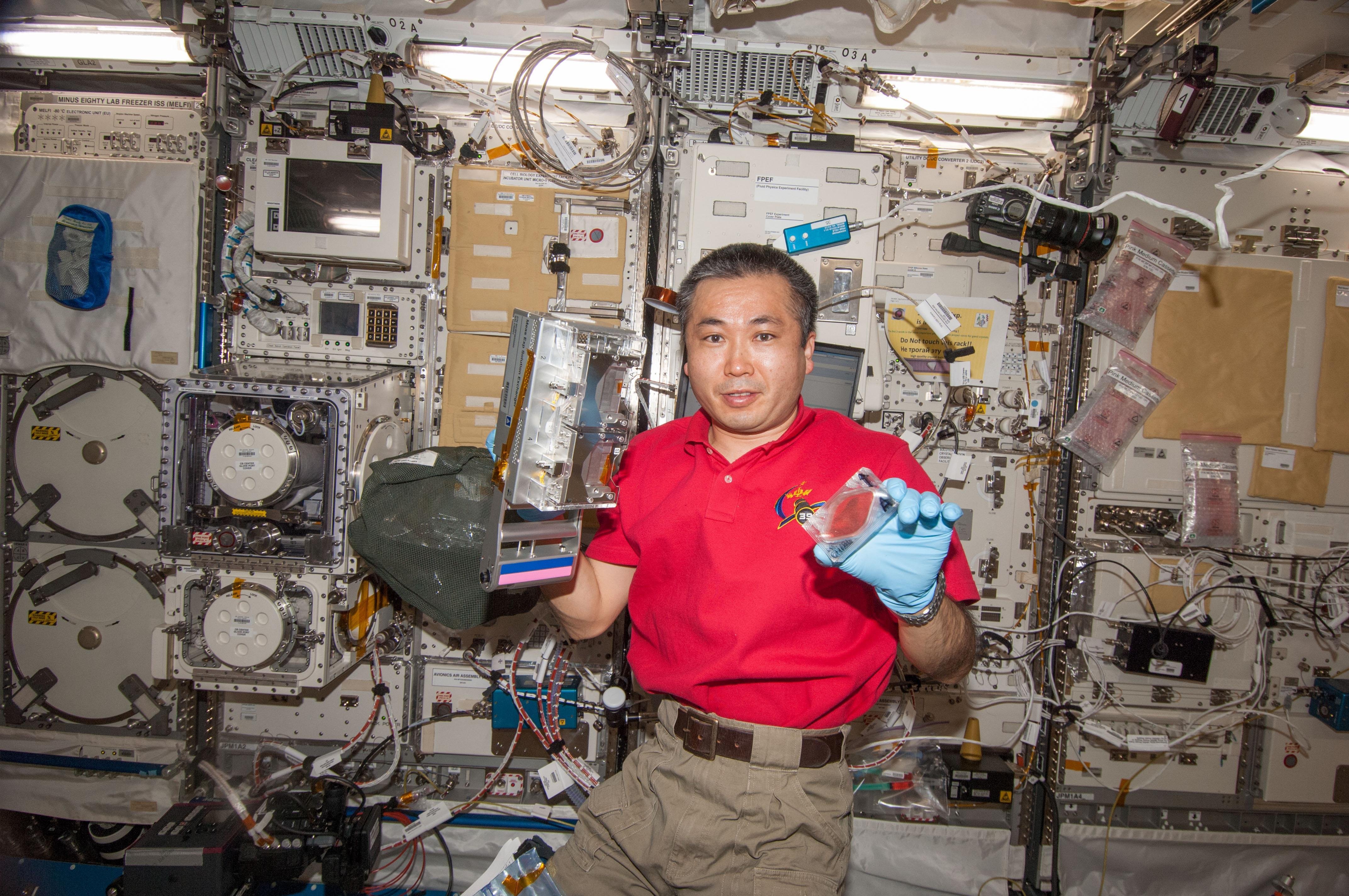
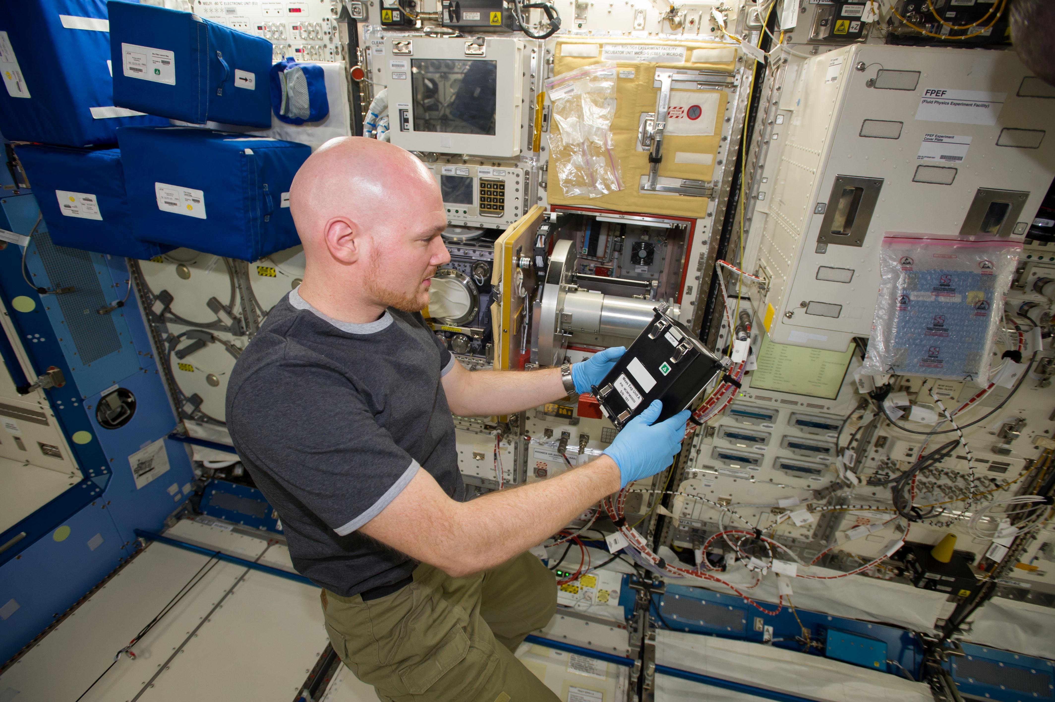
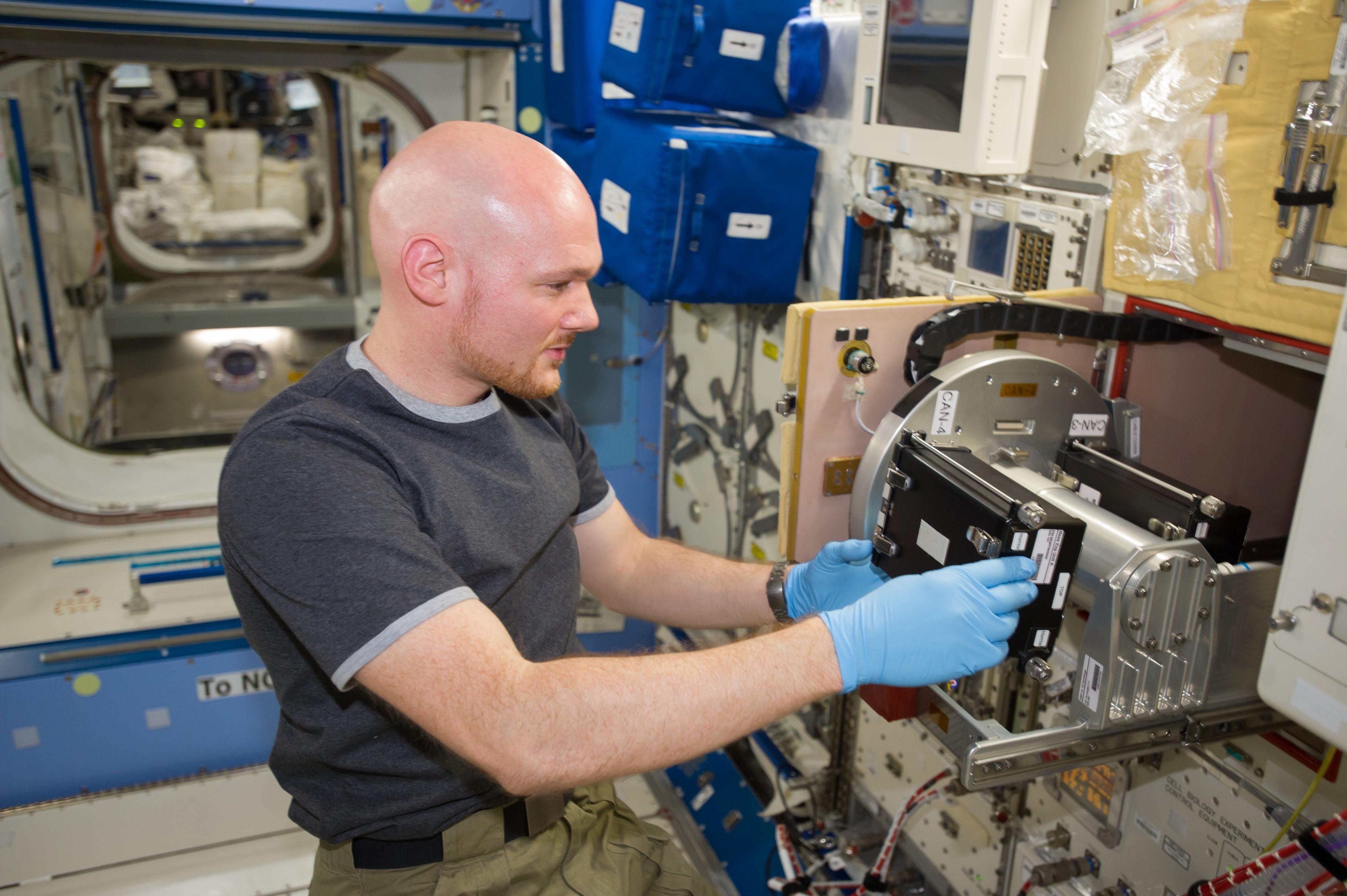
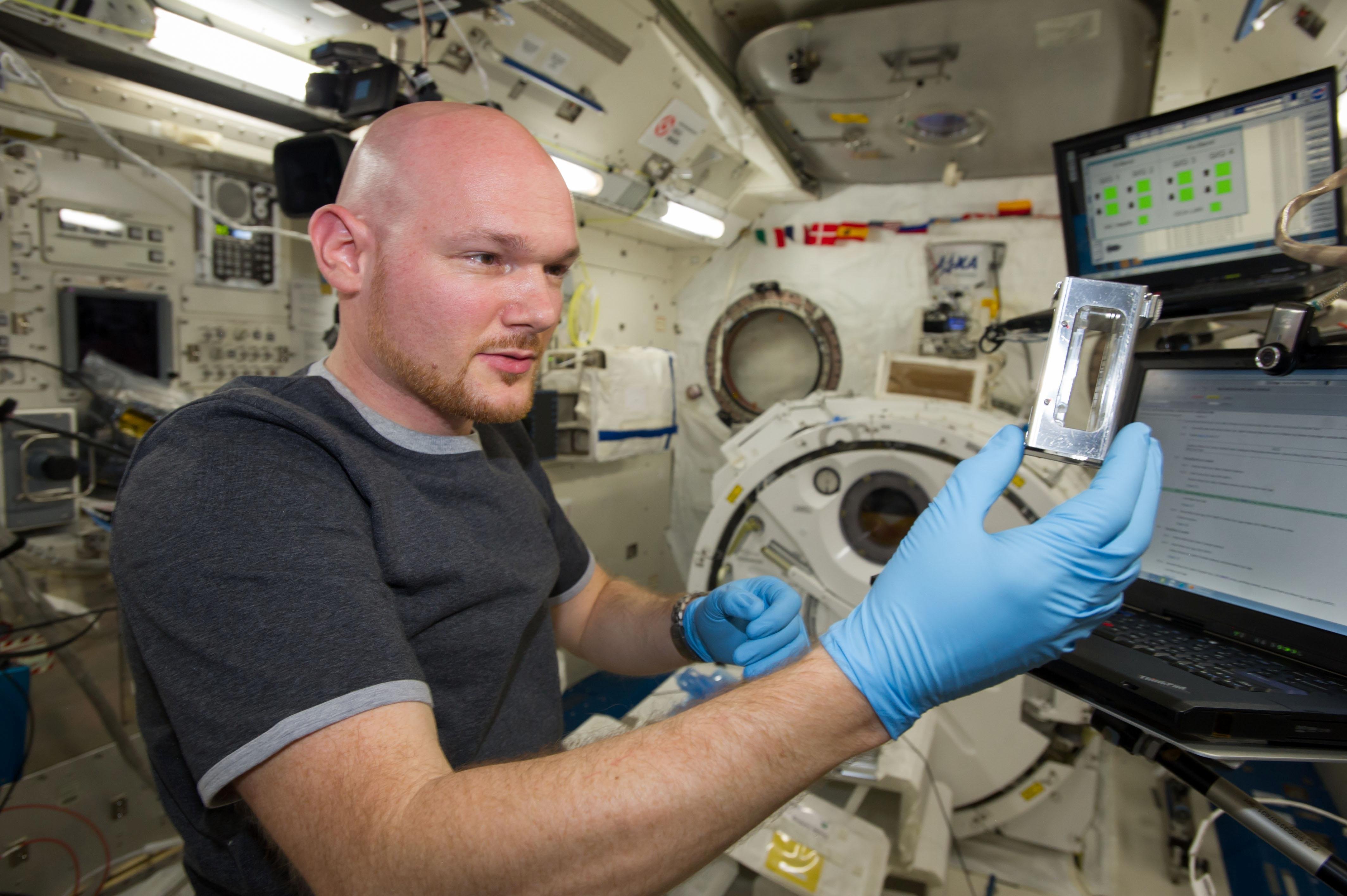
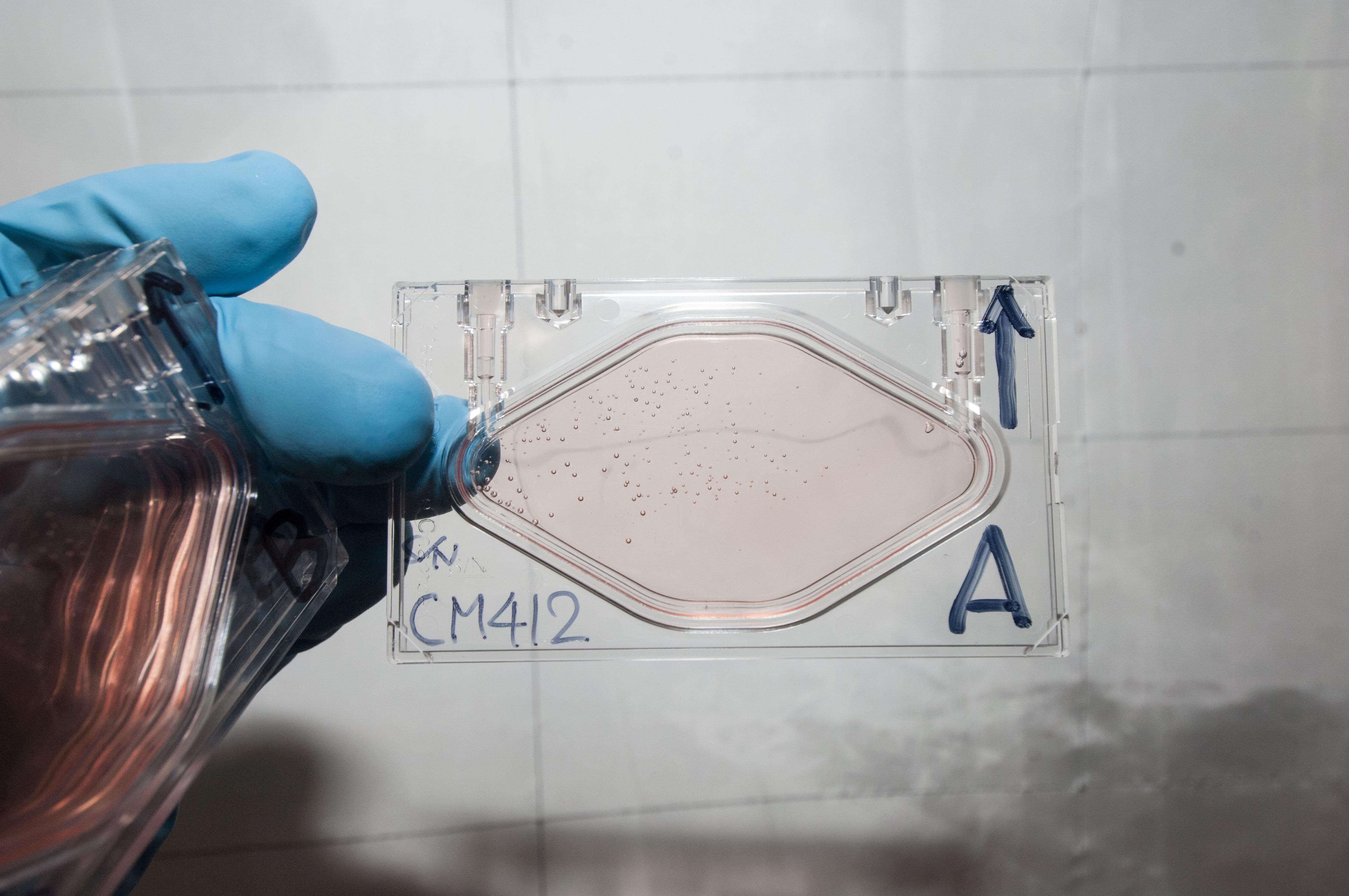
JAXA CBEF, Measurement Experiment Culture Chambers and Measurement Experiment Cages are used for incubation. Measurement Experiment Solution Exchangers are used for medium/fixative exchange.
Media Gallery

Applications
SPACE APPLICATIONS
Loss of muscle strength and tone, and impaired recovery after return to Earth, are long-standing space medicine challenges that resist exercise and other counter-measures in microgravity. Investigations like Cell Mechanosensing can help reveal the mechanical and chemical links that alter normal cell functions, and cause muscle atrophy, so researchers can develop inhibitors to combat microgravity-induced muscle atrophy.
EARTH APPLICATIONS
People subjected to long periods of bed rest and inactivity, such as the elderly or patients after surgery, experience severe muscle atrophy that is difficult to reverse. Some forms of muscular dystrophy also appear to parallel aspects of muscle wasting in space. Understanding the biochemical mechanisms at work will lead to improved therapeutic approaches for these disabilities.
Operations
OPERATIONAL REQUIREMENTS AND PROTOCOLS
The samples (L6 cells and A-6 cells) are incubated in JAXA CBEF with micro-gravity and artificial 1g environment. L6 cells and A-6 cells are cultured in Measurement Experiment/ Cell Experiment Culture Chambers in CBEF. L6 and A-6 cells are observed using fluorescence microscopy and downlinked images. Lastly, the cells are fixed with Paraformaldehyde for recovery (at 2°C).
Active L6 cells and A-6 cells are launched at ambient temperature, and then incubation in CBEF is started soon after docking. After 3 days of incubation, 2 chambers are observed, and cells are fixed using Cell Experiment Fixation Cylinder, then stored at 2°C in MELFI. Other chambers are continuously observed over the next 4 days.
Publications
PRINCIPAL INVESTIGATOR(S)
SOKABE Masahiro [Nagoya University]
Unless specified otherwise, rights to all images belong to ©JAXA